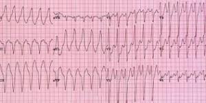
Ventricular Fibrillation Complicating Acute Myocardial Infarction
 Itoo Distinct Clinical and Electrocardiographic Features
Ventricular fibrillation (VF) is the major cause of cardiac death within the early hours of evolving infarction or ischemia. A number of clinical studies have attempted so far to evaluate the prognostic significance of primary VF complicating acute myocardial infarction (AMI), the role of sympathetic activity, the tendency for recurrence, and prophylactic treatment. Numerous studies have been designed in an attempt to define the predictive values of predisposing factors, such as the infarcts site and size, abnormalities of heart rate and conduction, prolonged corrected Q-T interval (Q-Tc), R-on-T phenomenon, warning arrhythmias, and metabolic disorders. None of these studies has commented on significant variability of fibrillation amplitude and configuration and their interpretation in relation to various stages of infarction. add comment
Primary VF has been defined when occurring without evidence of hypotension or heart failure during the first day of AMI or as primary electrical failure not associated with AMI. The purpose of this study was to characterize two distinct electrocardiographic patterns of primary VF complicating AMI and to correlate their course of appearance with specific electrocardiographic stages of evolving infarction. Different underlying mechanisms might be responsible for these two electrocardiographic patterns of primary VF.
Materials and Methods
A retrospective study was done of 69 consecutive patients admitted to our coronary care unit within two hours after the onset of symptoms, who developed VF in the setting of AMI within 24 hours from initial cardiac symptoms. The criteria for the diagnosis of infarction were severe retrosternal chest pain of more than 30 minutes’ duration, characteristic evolving electrocardiographic changes with the development of new pathologic Q waves, and typical elevation of serum cardiac enzymes compatible with AMI. A 12-lead electrocardiogram was recorded on the patients presentation to the emergency department and on arrival in the coronary care unit. Repeated ECGs taken consecutively at least every hour until two hours after the onset of VF were available. All patients had continuous electrocardiographic monitoring and the presence of VF defined as irregular, fast ventricular activity (>300/min) associated with total pump failure demonstrated by automatically recorded rhythm strips with identical positions of the electrodes in all patients.
The electrocardiographic evolution of AMI was divided as follows into four stages: (1) stage 1 consists of ST-segment elevation, peaked positive T waves, and Q waves of less than 2 mm; (2) stage 2 consists of Q waves of more than 2 mm with decrease in the size of R waves; ST segment is still elevated with positive T waves; (3) stage 3 consists of inversion of the previously positive T waves and still with ST-segment elevation; and (4) stage 4 consists of the ST segments return to the isoelectric baseline. A patients stage of AMI was defined by two observers who were blinded, using the ECG prior to the onset of VF.
Category: Myocardial Infarction
Tags: electrocardiographic, myocardial infarction, ventricular fibrillation