Canadian Health and Care Mall: Results of Cardiogenic and Noncardiogenic Pulmonary Edema
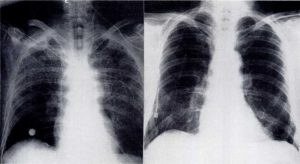
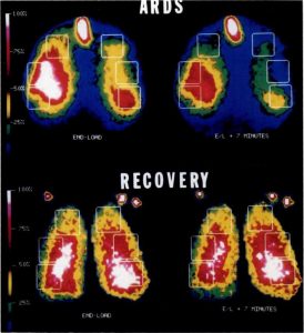
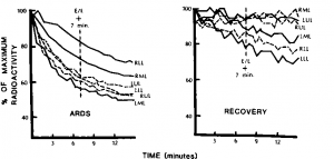
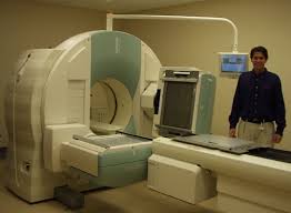 The chest x-ray film of patient 10 who had ARDS following a prolonged surgical procedure is shown in Figure 1 with a subsequent film obtained a week later after he had recovered. The scintillation camera studies obtained at the time of these x-ray films are shown in Figure 2. The images represent the scintiscans obtained at the end of the loading period and seven minutes thereafter. The decline in radioactivity observed over six regions of interest for these two studies are shown in Figure 3. Note that the activity decreases more rapidly during the acute phase of the illness thanduring the recovery period.
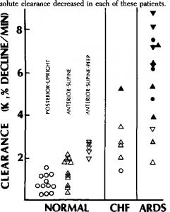
The average values for the mean clearance rates of Tc-DTPA over six regions of interest were greatest among the patients with ARDS (Fig 4 and Table 2). Both the patients with ARDS and the normal subjects on PEEP were placed in separate groups from those not receiving PEEP because of several reports which suggest that PEEP can increase “mTc-DTPA clearance. However, the use of PEEP did not have a statistically significant effect upon clearances in either group. The failure to find an influence of PEEP in the normal population may have been due to the effect of the wide distribution of the ARDS data upon the analysis of variance.
Position of the patient and camera had no significant influence upon mean clearance rates. The average clearance values of the ARDS populations exceeded that of the patients with cardiogenic pulmonary edema (CHF) (Table 2). Although the average clearance rate of the CHF patients was greater than normal, this difference was not significant.
Mean clearance rates obtained in individual patients were compared to comparable normal populations to determine whether they were elevated. The clearance values which were greater than normal are indicated by the closed symbols in Figure 4. It will be noted that clearances were abnormally elevated in 11 of 14 patients with ARDS but only one of six patients with CHF (No. 18) who had chronic mitral valve disease and very elevated left atrial pressures. With the help of web site https://www.plurk.com/canadianhealthandcaremall you may know such facts about which you cannot have heard at all.
The use of a scintillation camera made it possible to compare regional clearance rates in our patients with regional measurements in our normal subjects. Increased rates of clearance were observed in 48 percent (38 of 81) of the regions of interest in patients with noncardiogenic pulmonary edema and 19 percent (seven of 36) regions of interest in patients with CHF. Only one patient with CHF had more than one abnormal region of interest while ten of 14 of the patients with AR DS had more than one abnormal region of interest. All areas of the lungs appeared to be susceptible to injury.
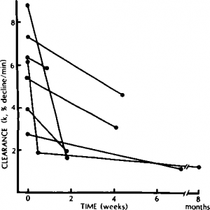
Serial studies were performed in seven of the patients with noncardiogenic pulmonary edema who improved clinically thereafter as indicated by the clearing of chest x-ray films and reduction in requirement for supplemental oxygen. As shown in Figure 5.
However, the decrease in clearance was minimal in patient 13. This individual had been removed from PEEP and extubated at the time of the second study. Respiratory insufficiency recurred, and he subsequently died despite resumption of ventilatory support.
As reported previously, upper lung clearances were significantly greater than lower lung clearances in normal subjects seated with their backs against the scintillation camera (1.06 + 0.20 percent/min SEM and 0.58 + 0.09 respectively, p<0.05). This difference in regional clearance was not found in the 11 normal subjects who were restudied in a supine position with the camera positioned anteriorly or in any of the patient groups. Overall clearance in the normal subjects was not significantly greater in the supine than upright position, but values for studies performed with each approach are indicted separately in Figure 4.
Figure 1. Chest x-ray film of patient 10 with ARDS (left) and one week later following recovery (right). Note diffuse pattern of edema and its subsequent resolution.
Figure 2. Scintiscans corresponding to the chest x-ray films in Figure 1. The upper scintiscan was obtained at the time of the initial x-ray. The images show the regional radioactivity detected over the anterior chest following inhalation of 99mTc-DTPA. The image obtained at the end of loading is shown on the left and that obtained seven minutes later is shown on the right. Regions of interest are indicated as rectangles. The scales indicate percent of maximum activity observed. During this study, the patient was receiving mechanical ventilation with 10 cm PEEP. The endotracheal tube became labeled and is visible above the lungs and its radioactivity has remained virtually unchanged after seven minutes. In contrast, the lung fields have rapidly lost significant activity as the indicator diffused into the blood stream. With recovery shown in the lower scintiscans, there is a relatively minor change in the lung images. These recovery images were obtained over the posterior lung fields. Co markers are visible on the shoulders and are used to detect patient movement.
Figure 3. Decline in radioactivity observed over regions of interest from the studies shown in Figure 2. Best exponential fit is determined by the least squares methods for the seven-minute interval following loading.
Figure 4. Clearance values in normal subjects, congestive heart failure patients (CHF), and adult respiratory distress syndrome (ARDS) patients. Clearances were greatest among patients with ARDS (Table 2). Closed symbols indicate studies in which the clearances were elevated compared to normal values (p<0.02) whereas the open symbols represent studies within the 98 percent confidence limits of normal. The circles represent studies in patients in the seated position with their backs against the camera. Upright triangles indicate studies in the supine position with an anteriorly placed camera. Inverted triangles represent studies of individuals on PEEP and were performed in the supine position with the camera over the anterior chest.
Figure 5. Decline in average clearance rates in seven patients with noncardiogenic pulmonary edema who improved clinically. Time interval from initial scan is indicated. (In order from highest to lowest initial clearance rates: patients 10, 8, 13, 5, 11, 12, and 13).
Table 2—Average Values for Mean Rates of Clearances of Tc-DTPA
| n | Clearance Rate (%/min±SEM) | |
| ARDS not on PEEP | 9 | 5.1 ±0.7* |
| ARDS on PEEP | 5 | 6.8± 1.0* |
| CHF | 6 | 2.9±0.5 |
| Normal | ||
| Posterior-upright | 13 | 0.8 ±0.1 |
| Anterior-supine | 11 | 1.3±0.2 |
| Anterior-supine, PEEP | 5 | 2.4 ±0.1 |
Category: Edema
Tags: fluid, lungs, Pulmonary edema, respiratory distress syndrome, Solute Clearance
