Restricted Pulmonary Function in Cystic Fibrosis: Discussion
 The results of this study indicate that restricted pulmonary function may be more common than previously reported in patients with cystic fibrosis. Among 158 patients, we identified ten with restriction (6 percent prevalence). Otherwise, restricted patients were remarkably similar to the matched control group of nonrestricted patients with respect to clinical and chest radiographic ratings. The restrictive defect did not appear to represent a more severe stage of disease, as has been proposed by others. Results of pulmonary function tests revealed significant differences between groups only for TLC(P), which was used in selecting restricted patients. The TLC(R) and TLC(N) were not significantly different between groups; however, nitrogen washout volume was performed in only five matched pairs. There were no differences in measurements of expiratory flow, Raw, maximal inspiratory pressure, or VC (reduced in both groups). read only
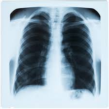
The discrepancy between groups for pulmonary volumes measured by the three different techniques highlights the important differences among these methods. Although the three methods produce equivalent results in normal subjects, each method measures a different theoretic volume which may differ with disease. Gas dilution techniques (nitrogen washout or helium dilution) measure communicating gas volume, the volume of gas which mixes with the reference gas. Body plethysmography measures compressible gas volume, the total volume of intrathoracic gas. This includes noncommunicating air space not measured by gas dilution or washout methods. Radiographic techniques estimate intrathoracic volume from the area bounded by the thoracic cage, diaphragm, and mediastinum.
In obstructive disease with noncommunicating air space, gas dilution measurements may underestimate both radiographic and plethysmographic volumes. In restrictive processes in which gas is replaced by fluid or abnormal tissue (eg, edema, fibrosis, pneumonitis), the radiographic thoracic volume may be larger than the gas dilution or plethysmographic volume. In restrictive conditions which do not affect the pulmonary parenchyma directly (eg, respiratory muscle weakness), the three techniques should be comparable.
Category: Pulmonary Function
Tags: airway, cystic fibrosis, Pulmonary Function