Canadian Health and Care Mall: Results of Electrocardiographic and Vectorcardiographic Diagnosis of Posterior Wall Myocardial Infarction
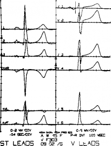
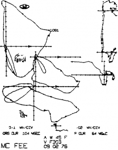
 Table 1 displays the individual angiographic characteristics of the 27 patients. Twenty-two had total and five had subtotal occlusion of the circumflex or obtuse example of an ECG and vectorcardiogram is shown in Figures 1 and 2.
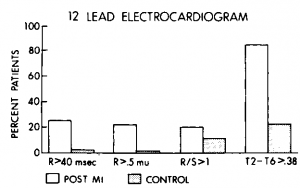
The electrocardiographic analysis is shown in Table 2. The total QRS duration was within normal limits. Pathologic Q waves of 35 msec or more in lead aVF were present in six patients (22 percent). The R wave in lead Vj was equal to or greater than 40 msec in seven patients (26 percent) and in lead V2 was equal to or greater than 50 msec in five patients (19 percent). The amplitude of the R wave in lead Vx was equal to or greater than 0.5 mV in six patients (22 percent), and the mean R/S ratio in leads Vx and V2 was 0.66 and 1.45, respectively. In lead Vx the ratio was greater than 1 in only six patients, and in lead V2, it was greater than 1.5 also in six patients (22 percent). With the exception of the QRS duration, all of these variables are significantly different from controls.
The T-wave voltage in lead V2 was 0.63 ±0.32 mV and in lead V6 was 0.08 ± 0.14 mV. In the control group, these values were 0.52 ±0.19 mV (p<0.05) and 0.34±0.16 mV (p<0.001), respectively.
The horizontal T angle as approximated by the T2 – T6 index was 0.55 ±0.35 mV in the group with infarction and 0.22 ± 0.15 mV in the control group (p<0.001). The true-positive ratio and the false-positive ratio of the T2 — Te index varying from 0.25 to 0.60 mV is shown in Table 3. A T2 — Te index greater than 0.38 mV appears to be the best discriminator, since it was found in 81 percent (22) of the patients and only 25 percent (24) of the controls. The sensitivity of selected electrocardiographic criteria is illustrated in Figure 3.
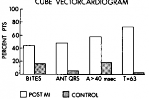
The vectorcardiographic characteristics are shown in Table 4 and Figure 4 (Cube) and in Table 5 and Figure 5 (McFee). The duration of the anterior portion of the QRS complex was 42 msec for the Cube and 46.4 msec for the McFee, both significantly different from the control group (p<0.001). In the Cube system the 40-msec vector was anterior to the E point in 59 percent (16) of the cases with myocardial infarction and in 20 percent (19) of the controls (p<0.05); in the McFee system, this vector was anterior in 85 percent (23) of the patients and in 45 percent (43) of the controls (not significant). The anterior voltage, 0.21 mV for the Cube and 0.65 mV for the McFee system, was similarly significantly greater than in the controls (p<0.01). The maximum posterior voltage was decreased in the patients with infarction (p<0.001). This resulted in a greater A/P ratio for the patients (2.1 in Cube and 1.8 in McFee) as compared to controls (p<0.002).
Approximately one-half of the patients (48 percent Cube and 52 percent McFee) had predominantly anterior QRS loops, while in the control group, this was present in less than 10 percent of the cases (p<0.001). To read more about erectile dysfunction you may on the LiveJournal official group with hot news from canadian healthcare mall.
The maximum leftward voltage was markedly decreased in the patients (0.40 and 1.16 mV for Cube and McFee, respectively) and significantly different from the controls (p<0.001).
The maximum T-vector angle in the horizontal plane was 72.2°±48.3° (Cube) and 66.3°±43.3° (McFee), compared to 24.2° ±19.4° (Cube) and 42.4° ±12.7° (McFee) for the control group (p<0.001).
Twenty of the 27 patients had a T-wave angle greater than 63° in the Cube, and 19 of them had an angle greater than 68° in the McFee, compared with 3 out of 97 of the control group (p<0.001). (The values 63 and 68 were obtained from the mean ± 2 SD in the normal population.
An analysis of the true-positive and false-positive ratio for the horizontal T angle is shown in Table 6 for both Cube and McFee vectorcardiographic lead systems.
In the Cube lead system, late QRS bites were (bund in 44 percent (12) of patients with infarction and 18 percent (17) of controls (p<0.01). In the McFee system, bites were present in 11 percent (three) of the group with infarction and 4 percent (4) of the controls (not significant).
Figure 1. Electrocardiogram of patient 13. Left ventricular angiogram showed 4-cm posterior wall infarction due to complete occlusion of large marginal branch. There was minimal disease in left anterior descending artery; right coronary artery was normal.
Figure 2. McFee vectorcardiogram of patient 13.
Figure 3. True-positive ratio and false-positive ratio of common electrocardiographic criteria for posterior wall infarction. MI, Myocardial infarction. R>40 msec=R wave>40 msec in lead R>.5 mV = R wave>0.5 mV in lead V,; R/S>1 = R/S ratio>l in lead Vt; and T2 — T6>. 38 = difference in T-wave voltages in leads V2 and Ve.
Figure 4. True positive ratio and false-positive ratio of Cube vectorcardiographic criteria for posterior wall infarction. MI, Myocardial infarction. ANT QRS = 50 percent loop anterior to E point; A>40 msec = 40-msec vector anterior to E point; and T>63 = T-wave angle greater than 63°.
Figure 5. True-positive ratio and false-positive ratio of McFee vectorcardiographic criteria for posterior wall infarction. MI, Myocardial infarction. ANT QRS = ^50 percent loop anterior to E point; A>40 msec=40-msec vector anterior to E point; and T>68 = T-wave angle greater than 68°.
Table 1—Angiographic Characteristics of Posterior Wall Infarction
| Case, Sex, Age (yr) | Percent of Occlusion* | Size of Infarct, cm | EjectionFraction,percent | ||
| LAD | CX | RCA | |||
| 1,M,35 | 0 | 100 | 0 | 2 | 60 |
| 2,M,54 | 0 | 100 | 0 | 2 | 64 |
| 3,M,51 | 50 | 100 | 65 | 2 | 61 |
| 4,F,42 | 0 | 95 | 0 | 3 | 59 |
| 5,M,60 | 0 | 100 | 0 | 3 | 57 |
| 6,M,44 | 70 | 100 | 65 | 4 | 63 |
| 7,M,42 | 0 | 100 | 0 | 3 | 61 |
| 8,M,42 | 50 | 100 | 60 | 3 | 64 |
| 9,M,52 | 70 | 100 | 70 | 4 | 58 |
| 10,M,48 | 70 | 100 | 40 | 4 | 48 |
| 11,M,55 | 0 | 100 | 50 | 5 | 58 |
| 12,M,49 | 0 | 100 | 65 | 4 | 53 |
| 13,F,45 | 40 | 100 | 0 | 4 | 53 |
| 14,M,50 | 0 | 100 | 20 | 5 | 54 |
| 15,M,59 | 65 | 100 | 50 | 5 | 49 |
| 16,M,54 | 0 | 99 | 0 | 5 | 44 |
| 17,M,54 | 50 | 100 | 0 | 6 | 51 |
| 18,M,50 | 0 | 100 | 70 | 6 | 48 |
| 19,M,49 | 0 | 100 | 0 | 3 | 61 |
| 20,M,74 | 50 | 100 | 0 | 6 | 51 |
| 21,M,46 | 70 | 100 | 0 | 7 | 43 |
| 22,M,52 | 70 | 95 | 0 | 4 | 62 |
| 23,M,50 | 40 | 100 | 0 | 8 | 44 |
| 24,M,58 | 65 | 100 | 0 | 5 | 48 |
| 25,M,38 | 65 | 100 | 30 | 2 | 70 |
| 26,M,40 | 70 | 95 | 0 | 6 | 47 |
| 27,M,50 | 0 | 90 | 0 | 2 | 67 |
Table 2—Electrocardiographic Analysis of Posterior Wall Infarction
| Case1 | QRSDur,msec*
116 |
Q-Wav<iLead
2 30 |
г Dun msecLead3
35 |
ition,LeadaVF
35 |
R-WDurams
Lead v, 31 |
/aveition,ec
Lead v2 39 |
R-WAmplim
Lead v, 0.2 |
/aveitude,V
Lead v2 0.7 |
S-VAmplim
Lead Vi 0.5 |
/aveitude,V
Lead v2 1.4 |
R/S ILeadv,
0.40 |
tatioLeadv2
0.50 |
T-WavLeadv2
0.80 |
re Ampli mVLeadv6 ’ 0.10 | tude,v2-v6t0.70 | Notev, | :h Rv2 | Notv, | ch Sv2 |
| 2 | 106 | 0 | 0 | 0 | 35 | 40 | 0.3 | 0.5 | 1.3 | 0.5 | 0.23 | 1.00 | 1.00 | 0.25 | 0.75 | – | – | – | – |
| 3 | 92 | 0 | 0 | 0 | 23 | 20 | 0.2 | 0.3 | 0.4 | 0.8 | 0.50 | 0.37 | 0.50 | 0.15 | 0.35 | – | – | + | + |
| 4 | 90 | 20 | 20 | 20 | 30 | 40 | 0.2 | 0.6 | 0.2 | 0.4 | 1.00 | 1.50 | 0.70 | 0.10 | 0.60 | – | – | – | – |
| 5 | 82 | 20 | 0 | 0 | 27 | 50 | 0.2 | 0.5 | 0.5 | 0.6 | 0.40 | 0.83 | 0.60 | 0.15 | 0.45 | – | + | – | – |
| 6 | 96 | 0 | 0 | 0 | 31 | 43 | 0.2 | 0.9 | 0.2 | 0.6 | 1.00 | 1.50 | 0.70 | 0.30 | 0.40 | – | – | + | + |
| 7 | 100 | 25 | 35 | 25 | 35 | 35 | 0.3 | 0.6 | 0.8 | 0.8 | 0.37 | 0.75 | 0.90 | 0.10 | 0.80 | – | – | – | – |
| 8 | 90 | 0 | 20 | 0 | 30 | 35 | 0.4 | 0.7 | 0.7 | 0.9 | 0.57 | 0.77 | 0.40 | -0.10 | 0.50 | – | – | – | – |
| 9 | 90 | 30 | 0 | 20 | 30 | 30 | 0.5 | 0.5 | 0.8 | 1.2 | 0.62 | 0.41 | 0.60 | 0.05 | 0.55 | + | + | – | – |
| 10 | 90 | 0 | 0 | 0 | 40 | 47 | 0.8 | 1.5 | 0.8 | 1.2 | 1.00 | 1.25 | 1.25 | 0.15 | 1.10 | – | – | – | – |
| 11 | 90 | 35 | 50 | 40 | 40 | 40 | 0.5 | 1.4 | 0.9 | 1.2 | 0.55 | 1.16 | 0.55 | 0.05 | 0.50 | – | – | – | – |
| 12 | 84 | 30 | 35 | 30 | 23 | 27 | 0.6 | 0.8 | 0.7 | 1.3 | 0.85 | 0.61 | 0.70 | 0.10 | 0.60 | – | – | + | – |
| 13 | 100 | 20 | 25 | 20 | 25 | 30 | 0.3 | 0.7 | 0.8 | 0.8 | 0.37 | 0.87 | 0.85 | 0.30 | 0.55 | – | – | + | + |
| 14 | 106 | 30 | 40 | 40 | 30 | 35 | 0.2 | 0.3 | 0.3 | 0.5 | 0.66 | 0.60 | 0.55 | 0.25 | 0.30 | – | + | – | + |
| 15 | 90 | 25 | 25 | 25 | 40 | 40 | 0.4 | 0.8 | 0.6 | 0.7 | 0.66 | 1.14 | 0.95 | -0.25 | 1.20 | – | – | – | – |
| 16 | 110 | 20 | 50 | 80 | 50 | 50 | 0.4 | 0.8 | 0.2 | 0.6 | 2.00 | 1.33 | 0.40 | 0.00 | 0.40 | – | – | – | – |
| 17 | 110 | 20 | 0 | 0 | 35 | 40 | 0.3 | 0.4 | 1.0 | 1.1 | 0.30 | 0.36 | 0.65 | -0.20 | 0.85 | – | – | – | – |
| 18 | 80 | 0 | 10 | 10 | 25 | 25 | 0.2 | 0.2 | 0.8 | 1.1 | 0.25 | 0.18 | 0.55 | 0.00 | 0.55 | + | + | – | – |
| 19 | 95 | 25 | 0 | 20 | 40 | 50 | 0.4 | 1.3 | 0.5 | 0.5 | 0.80 | 2.60 | 0.70 | 0.15 | 0.55 | – | – | – | – |
| 20 | 100 | 25 | 20 | 20 | 40 | 40 | 0.2 | 0.7 | 0.4 | 1.1 | 0.50 | 0.63 | -0.20 | 0.25 | -0.45 | – | + | – | – |
| 21 | 80 | 30 | 0 | 45 | 40 | 50 | 0.9 | 1.3 | 0.9 | 0.4 | 1.00 | 3.25 | 0.90 | 0.05 | 0.85 | – | – | – | – |
| 22 | 95 | 25 | 0 | 0 | 31 | 43 | 0.1 | 0.7 | 0.4 | 0.8 | 0.25 | 0.87 | 0.60 | 0.15 | 0.45 | + | – | – | – |
| 23 | 92 | 40 | 50 | 70 | 30 | 50 | 0.3 | 1.0 | 0.4 | 0.2 | 0.75 | 5.00 | 0.80 | -0.15 | 0.95 | – | – | – | – |
| 24 | 105 | 30 | 30 | 30 | 30 | 40 | 0.6 | 2.0 | 1.0 | 1.0 | 0.60 | 2.00 | 0.35 | 0.00 | 0.35 | – | – | – | – |
| 25 | 78 | 0 | 0 | 0 | 15 | 45 | 0.1 | 0.9 | 0.7 | 0.1 | 0.14 | 9.00 | 0.80 | 0.15 | 0.65 | – | – | – | – |
| 26 | 90 | 30 | 0 | 30 | 30 | 40 | 0.4 | 0.5 | 0.2 | 1.0 | 2.00 | 0.50 | -0.30 | -0.05 | -0.35 | – | – | + | – |
| 27 | 80 | 25 | 35 | 30 | 20 | 20 | 0.1 | 0.1 | 0.8 | 0.9 | 0.12 | 0.11 | 0.80 | 0.10 | 0.70 | – | – | – | – |
| Mean | 94 | 20 | 18 | 22 | 32 | 39 | 0.3 | 0.8 | 0.6 | 0.8 | 0.66 | 1.45 | 0.63 | 0.08 | 0.55 | 3 | 5 | 5 | 4 |
| SD | 10 | 13 | 19 | 21 | 8 | 9 | 0.2 | 0.4 | 0.3 | 0.3 | 0.47 | 1.83 | 0.32 | 0.14 | 0.35 | ||||
| nControl | 27 | 27 | 27 | 27 | 27 | 37 | 27 | 27 | 27 | 27 | 27 | 27 | 27 | 27 | 27 | ||||
| Mean | 94 | 10 | 10 | 10 | 25 | 31 | 0.2 | 0.5 | 0.8 | 1.2 | 0.28 | 0.48 | 0.52 | 0.34 | 0.22 | 3 | 8 | 11 | 4 |
| SD | 9 | 10 | 12 | 10 | 7 | 9 | 0.1 | 0.2 | 0.28 | 0.4 | 0.19 | 0.27 | 0.19 | 0.16 | 0.15 | ||||
| n | 97 | 97 | 97 | 97 | 97 | 97 | 97 | 97 | 97 | 97 | 97 | 97 | 97 | 97 | 97 | ||||
| t | 0.05 | 4.08 | 2.65 | 4.02 | 4.66 | 3.67 | 5.02 | 4.39 | 3.09 | 4.57 | 6.35 | 5.07 | 2.25 | 7.6 | 7.2 | ||||
| x2 | 1.4 | 1.4 | 0.4 | 2.42 | |||||||||||||||
| P | NS$ | <0.001 | 0.01 | 0.001 | 0.001 | 0.001 | 0.001 | 0.001 | 0.005 | 0.001 | 0.001 | 0.001 | 0.05 | 0.001 | 0.001 | NS | NS | NS | NS |
Table 3—T2 — Te Index in Posterior Wall Infarction
| T2-Te | PercentTrue-Positive | PercentFalse-Positive |
| >0.25 | 93 | 49 |
| 0.30 | 89 | 38 |
| 0.35 | 81 | 29 |
| 0.38 | 81 | 25 |
| 0.40 | 74 | 21 |
| 0.45 | 66 | 15 |
| 0.50 | 60 | 11 |
| 0.60 | 37 | 4 |
Table 4—Vectorcardiographic Analysis with Cube Lead System
| Duration, msec | Voltage, mV | T-WaveAngle,degrees | Voltage Ratios | 50PercentAnt | ||||||||
| Case | I Ant | T Post | Ant | Leftward | Post | I Right | Bites | Left/I Right Left/Ant | Ant/Post | |||
| 1 | 43 | 57 | 0.11 | 0.21 | 0.24 | 0.01 | 35 | – | 21.00 | 1.90 | 0.46 | – |
| 2 | 33 | 61 | 0.17 | 0.80 | 0.52 | 0.13 | 55 | – | 6.15 | 4.70 | 0.32 | – |
| 3 | 27 | 63 | 0.13 | 0.20 | 0.17 | 0.04 | 50 | + | 5.00 | 1.53 | 0.76 | – |
| 4 | 48 | 42 | 0.18 | 0.27 | 0.25 | 0.05 | 100 | + | 5.40 | 1.50 | 0.72 | + |
| 5 | 34 | 44 | 0.16 | 0.67 | 0.18 | 0.19 | 70 | – | 3.52 | 4.18 | 0.89 | – |
| 6 | 42 | 10 | 0.11 | 0.45 | 0.02 | 0.06 | 5 | + | 7.50 | 4.09 | 5.50 | + |
| 7 | 36 | 44 | 0.11 | 0.52 | 0.20 | 0.04 | 105 | – | 13.00 | 4.72 | 0.50 | – |
| 8 | 42 | 46 | 0.32 | 0.37 | 0.33 | 0.02 | 115 | – | 18.50 | 1.15 | 0.97 | + |
| 9 | 40 | 40 | 0.27 | 0.25 | 0.34 | 0.14 | 90 | – | 1.78 | 0.92 | 0.79 | + |
| 10 | 46 | 36 | 0.23 | 0.69 | 0.28 | 0.11 | 75 | – | 6.20 | 3.00 | 0.82 | + |
| 11 | 45 | 47 | 0.40 | 0.31 | 0.56 | 0.11 | 90 | – | 2.82 | 0.77 | 0.71 | – |
| 12 | 27 | 33 | 0.37 | 0.41 | 0.32 | 0.15 | 80 | + | 2.70 | 1.11 | 1.15 | + |
| 13 | 34 | 44 | 0.16 | 0.69 | 0.21 | 0.03 | 40 | – | 23.00 | 4.30 | 0.76 | – |
| 14 | 40 | 10 | 0.13 | 0.39 | 0.04 | 0.04 | 70 | + | 9.75 | 3.00 | 3.25 | + |
| 15 | 39 | 51 | 0.28 | 0.21 | 0.32 | 0.08 | 120 | + | 2.62 | 0.75 | 0.87 | – |
| 16 | 69 | 41 | 0.27 | 0.10 | 0.08 | 0.05 | 110 | + | 2.00 | 0.37 | 3.37 | + |
| 17 | 45 | 55 | 0.15 | 0.72 | 0.31 | 0.12 | 140 | – | 6.00 | 4.80 | 0.48 | – |
| 18 | 35 | 30 | 0.27 | 0.14 | 0.27 | 0.01 | 100 | – | 14.00 | 0.52 | 1.00 | – |
| 19 | 39 | 52 | 0.22 | 0.35 | 0.20 | 0.15 | 65 | + | 2.30 | 1.59 | 1.10 | + |
| 20 | 51 | 46 | 0.11 | 0.36 | 0.18 | 0.09 | 0 | + | 4.00 | 3.27 | 0.61 | + |
| 21 | 44 | 35 | 0.51 | 0.54 | 0.41 | 0.19 | 105 | – | 2.80 | 1.06 | 1.24 | + |
| 22 | 40 | 50 | 0.08 | 0.13 | 0.20 | 0.03 | 65 | – | 4.33 | 1.62 | 0.40 | – |
| 23 | 92 | 0 | 0.26 | 0.30 | 0.01 | 0.23 | 125 | + | 1.30 | 1.15 | 26.00 | + |
| 24 | 44 | 56 | 0.50 | 0.53 | 0.36 | 0.20 | 100 | + | 2.65 | 1.06 | 1.39 | + |
| 25 | 30 | 40 | 0.10 | 0.81 | 0.35 | 0.01 | 70 | – | 81.00 | 8.10 | 0.28 | – |
| 26 | 42 | 50 | 0.16 | 0.14 | 0.11 | 0.18 | -100 | + | 0.77 | 0.87 | 1.45 | – |
| 27 | 30 | 50 | 0.08 | 0.25 | 0.28 | 0.07 | 70 | – | 3.57 | 3.12 | 0.28 | – |
| Mean | 42 | 42 | 0.21 | 0.40 | 0.25 | 0.09 | 72 | 12/27 | 9.40 | 2.38 | 2.07 | 13/27 |
| SD | 13 | 15 | 0.12 | 0.21 | 0.13 | 0.07 | 48 | 15.50 | 1.88 | 4.91 | ||
| nControl | 27 | 27 | 27 | 27 | 27 | 27 | 27 | 27 | 27 | 27 | ||
| Mean | 35 | 58 | 0.16 | 0.68 | 0.40 | 0.05 | 24 | 17/95 | 19.40 | 5.56 | 0.47 | 6/95 |
| SD | 7 | 11 | 0.07 | 0.19 | 0.18 | 0.03 | 19 | 15.90 | 3.95 | 0.33 | ||
| n | 95 | 95 | 95 | 95 | 95 | 95 | 95 | 95 | 95 | 95 | ||
| t | 4.03 | 6.12 | 2.75 | 6.60 | 4.03 | 4.30 | 7.78 | 2.90 | 3.95 | 3.18 | ||
| x2 | 6.77 | 24.89 | ||||||||||
| P | 0.001 | 0.001 | 0.01 | 0.001 | 0.001 | 0.001 | 0.001 | 0.01 | 0.01 | 0.001 | 0.002 | 0.001 |
Table 5—Vectorcardiographic Analysis with McFee Lead System
| Duration, msec | Voltage, mV | T-WaveAngle,degrees | Voltage Ratios | 50 | ||||||||
| Case | I Ant | T Post | Ant | Leftward | Post | I Right | Bites | Left/I Right | Left/Ant | Ant/Post | PercentAnt | |
| 1 | 45 | 65 | 0.52 | 0.65 | 1.00 | 0.01 | 55 | – | 65.00 | 1.25 | 0.52 | – |
| 2 | 57 | 43 | 0.55 | 2.10 | 0.76 | 0.25 | 70 | – | 8.40 | 3.82 | 0.72 | – |
| 3 | 42 | 48 | 0.35 | 1.05 | 0.85 | 0.07 | 60 | – | 15.00 | 3.00 | 0.41 | – |
| 4 | 43 | 47 | 0.48 | 0.74 | 0.45 | 0.02 | 70 | – | 37.00 | 1.48 | 1.06 | + |
| 5 | 49 | 33 | 0.42 | 1.53 | 0.70 | 0.30 | 70 | – | 5.10 | 3.64 | 5.00 | – |
| 6 | 45 | 50 | 0.65 | 1.40 | 0.38 | 0.05 | 40 | – | 28.00 | 2.10 | 1.71 | + |
| 7 | 45 | 40 | 0.60 | 1.17 | 0.61 | 0.05 | 85 | – | 24.30 | 1.95 | 0.98 | – |
| 8 | 40 | 49 | 0.95 | 0.70 | 0.90 | 0.02 | 110 | – | 35.00 | 0.73 | 1.05 | + |
| 9 | 42 | 40 | 0.46 | 0.95 | 0.98 | 0.30 | 90 | – | 3.16 | 2.06 | 0.47 | – |
| 10 | 48 | 34 | 1.20 | 1.50 | 0.75 | 0.10 | 75 | – | 15.00 | 1.25 | 1.60 | + |
| 11 | 53 | 37 | 1.05 | 1.30 | 0.86 | 0.35 | 75 | – | 3.71 | 1.23 | 1.22 | + |
| 12 | 27 | 53 | 0.66 | 1.00 | 0.55 | 0.20 | 70 | + | 5.00 | 1.51 | 1.20 | – |
| 13 | 38 | 43 | 0.62 | 0.89 | 0.91 | 0.02 | 70 | – | 44.50 | 1.43 | 0.68 | – |
| 14 | 44 | 58 | 0.28 | 0.90 | 0.44 | 0.01 | 60 | – | 90.00 | 3.20 | 0.63 | – |
| 15 | 51 | 39 | 0.88 | 1.05 | 0.54 | 0.08 | 115 | – | 13.12 | 1.19 | 1.62 | + |
| 16 | 52 | 59 | 0.72 | 0.64 | 0.43 | 0.08 | 85 | – | 8.00 | 0.88 | 1.67 | + |
| 17 | 58 | 48 | 0.40 | 1.31 | 0.74 | 0.08 | 90 | – | 16.37 | 3.27 | 0.54 | — |
| 18 | 33 | 47 | 0.40 | 0.58 | 0.86 | 0.02 | 95 | – | 29.00 | 1.45 | 0.46 | – |
| 19 | 58 | 32 | 1.00 | 1.50 | 0.42 | 0.25 | 60 | – | 6.00 | 1.50 | 2.22 | + |
| 20 | 45 | 55 | 0.70 | 0.85 | 1.05 | 0.02 | -20 | – | 42.50 | 1.21 | 0.66 | + |
| 21 | 53 | 19 | 1.00 | 1.93 | 0.48 | 0.01 | 75 | – | 193.00 | 1.93 | 2.08 | + |
| 22 | 45 | 40 | 0.52 | 0.75 | 0.51 | 0.05 | 70 | – | 15.00 | 1.44 | 1.02 | + |
| 23 | 60 | 10 | 0.90 | 1.40 | 0.06 | 0.35 | 95 | – | 4.00 | 1.55 | 16.00 | + |
| 24 | 53 | 57 | 1.15 | 1.63 | 0.87 | 0.60 | 110 | + | 2.71 | 1.42 | 1.32 | + |
| 25 | 51 | 20 | 0.70 | 2.24 | 0.21 | 0.01 | 55 | – | 224.00 | 3.20 | 3.33 | + |
| 26 | 40 | 56 | 0.35 | 0.40 | 0.57 | 0.35 | -110 | + | 1.14 | 1.14 | 0.61 | – |
| 27 | 36 | 40 | 0.12 | 1.10 | 0.71 | 0.06 | 70 | – | 18.33 | 9.16 | 0.17 | – |
| Mean | 46 | 43 | 0.65 | 1.16 | 0.65 | 0.13 | 66 | 3/27 | 35.24 | 2.14 | 1.81 | 14/27 |
| SD | 8 | 12.9 | 0.28 | 0.47 | 0.25 | 0.15 | 44 | 54.24 | 1.65 | 3.01 | ||
| nControl | 27 | 27 | 27 | 27 | 27 | 27 | 27 | 27 | 27 | 27 | ||
| Mean | 38 | 56 | 0.44 | 1.50 | 0.81 | 0.08 | 42 | 4/95 | 41.2 | 3.86 | 0.61 | 9/95 |
| SD | 7 | 10 | 0.17 | 0.41 | 0.27 | 0.07 | 13 | 49.9 | 1.77 | 0.33 | ||
| n | 95 | 95 | 95 | 95 | 95 | 95 | 95 | 95 | 95 | 95 | ||
| t | 5.49 | 5.34 | 4.82 | 3.69 | 2.80 | 2.95 | 4.72 | 0.53 | 4.50 | 3.84 | ||
| x2 | 0.79 | 21.99 | ||||||||||
| P | 0.001 | 0.001 | 0.001 | 0.001 | 0.01 | 0.005 | 0.001 | NSt | NSt | 0.001 | 0.001 | 0.001 |
Table 6—Horizontal Т-Angle in Posterior Wall Infarction
| Horizontal Т-Wave Angle | Cube | McFee | ||
| PercentTrue-Positive | PercentFalse-Positive | PercentTrue-Positive | PercentFalse-Positive | |
| >55° | 74 | 5 | 81 | 10 |
| >60° | 74 | 3 | 70 | 3 |
| >63° | 74 | 3 | 70 | 3 |
| >65° | 66 | 3 | 70 | 3 |
| >68° | 59 | 3 | 70 | 3 |
| >70° | 52 | 3 | 44 | 0 |
Category: Infarction
Tags: cardiac catheterization, circumflex coronary artery, ventriculography, Wall Myocardial Infarction


