Posted in AIDS | 06/02/2015
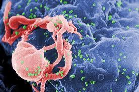
Pneumocystis carinii is a pathogen common to the respiratory tracts of immunosuppressed patients, which has recently become more prevalent with its frequent occurrence in the setting of the acquired immune deficiency syndrome (AIDS). Its typical radiographic presentation is that of diffuse bilateral, “fluffy” infiltrates on chest x-ray film. Histologically, this usually corresponds to a foamy, acellular eosinophilic intra-alveolar exudate in which the cysts of the organism are readily demonstrated with silver stains. The radiographic presentation of P carinii pneumonia as a single nodule has only rarely been reported,’ with some of the reports not having provided adequate descriptions of the lesions histologic appearances. website
Read more

Posted in Pulmonary Function | 05/02/2015
The results of serial pulmonary function tests from the previous (1983-84) and subsequent (1985-86) years also raises interesting questions about the mechanism of restriction and the implication of restriction regarding severity of disease. Three of nine restricted patients were no longer restricted on testing one year later, and two of eight were not restricted one year earlier. It is unlikely that the reduced plethysmographic pulmonary volume was due to measurement error, since nine of the ten restricted patients were restricted in at least two of the three years examined. In the nonrestricted patients, none of eight was restricted one year later, while two of eight were restricted one year earlier. Chest radiographic ratings did not change consistently over this period. Group data over one year of follow-up demonstrated a trend toward increase in expiratory flow and pulmonary volume in the restricted patients but not in the controls. These findings suggest…
Read more
Posted in Pulmonary Function | 03/02/2015
In this study, we proposed that restriction would be due to scarring and infiltration of the pulmonary parenchyma and, therefore, expected to find differences between the radiographic and gas volumes in the restricted patients. Although these differences did exist and, on average, were in the expected direction, there was considerable variability within groups, and the differences did not reach statistical significance. We also expected to find that the radiographic-gas volume differences in the restricted patients would correlate with radiographic grades of pulmonary parenchymal lesions (air-space or nodular cystic lesions) as a reflection of severity Surprisingly, the radio-graphic-gas volume differences were highly correlated only with radiographic ratings of air trapping (hyperinflation) and bronchial markings. These findings suggest that the mechanism of restriction in these patients with cystic fibrosis is not related directly to the extent of air-space-filling lesions and raises the interesting possibility that the restriction may be related more to…
Read more
Posted in Pulmonary Function | 02/02/2015
The results of this study indicate that restricted pulmonary function may be more common than previously reported in patients with cystic fibrosis. Among 158 patients, we identified ten with restriction (6 percent prevalence). Otherwise, restricted patients were remarkably similar to the matched control group of nonrestricted patients with respect to clinical and chest radiographic ratings. The restrictive defect did not appear to represent a more severe stage of disease, as has been proposed by others. Results of pulmonary function tests revealed significant differences between groups only for TLC(P), which was used in selecting restricted patients. The TLC(R) and TLC(N) were not significantly different between groups; however, nitrogen washout volume was performed in only five matched pairs. There were no differences in measurements of expiratory flow, Raw, maximal inspiratory pressure, or VC (reduced in both groups). read only
Read more
Posted in Pulmonary Function | 01/02/2015
Serial Studies Seventeen of the 20 patients (including seven matched pairs) had pulmonary function tests repeated during their 1985-86 annual examination, one year after the initial study. One restricted patient died prior to repeat assessment. Two nonrestricted patients did not have repeat tests (one due to an exacerbation of disease and one was lost to follow-up). Results of repeat pulmonary function tests in seven matched pairs showed a trend toward improvement in the restricted patients but not in the control subjects. The TLC increased 0.52 L (4.75 to 5.22 L) in the restricted and decreased 0.09 L (5.88 to 5.79 L) in the nonrestricted patients (p = 0.08 for time, p = 0.12 for group, and p = 0.02 for interactive effects). The VC increased an average of 0.45 L (2.63 to 3.08 L) in the restricted and decreased 0.19 L (2.62 to 2.43 L) in the nonrestricted patients (not…
Read more

Posted in Pulmonary Function | 27/01/2015
Pulmonary Function Tests General characteristics and results of selected pulmonary function tests of the restricted and matched nonrestricted patients are summarized in Table 1. There were no significant differences between groups for age, height, and weight, the variables used for matching. A significant difference was observed only for plethysmographic TLC (TLC[P]), which was used in the selection of restricted patients. Plethysmographic RV and FRC were lower in the restricted group but not significantly different from the nonrestricted patients. The TLC by nitrogen washout (TLC [N]) was measured in only six restricted and eight nonrestricted patients. In five pairs of matched subjects, TLC(N) was not significantly lower in the restricted group. Radiographic TLC (TLC[R]) was also not significantly different between the groups. In addition, there were no differences in VC or expiratory flow rates (reduced in both groups), Raw (elevated in both groups), maximal inspiratory pressure (normal), or change in FEVX…
Read more
Posted in Pulmonary Function | 26/01/2015
Pulmonary Function Tests Spirometry was performed on a spirometer (Ohio 842) according to recommendations of the American Thoracic Society’s Snowbird Conference before and after bronchodilator administration. Absolute pulmonary volumes were measured by three techniques: (1) body plethysmography using a constant-volume plethysmograph (Collins); (2) open-cireuit nitrogen washout; and (3) radiographic TLC by planimetry Plethysmographic airway resistance (Raw) and maximal inspiratory pressure were also measured. Reference values used were those of Knudson and co-workers for spirometry and those of Polgar and Promadhat (age less than or equal to 16) or Naimark and co-workers (age over 16) for pulmonary volumes. type two diabetes medications
Read more